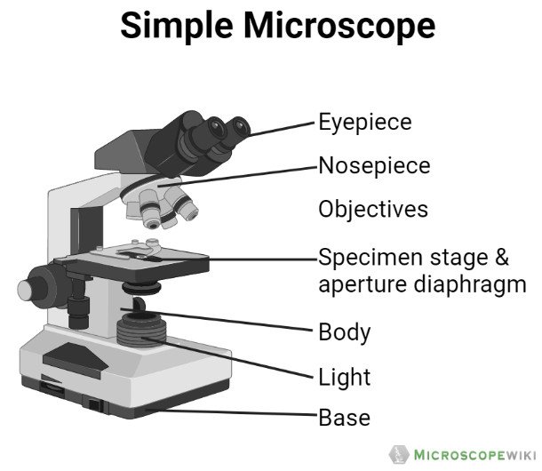
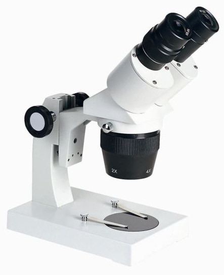
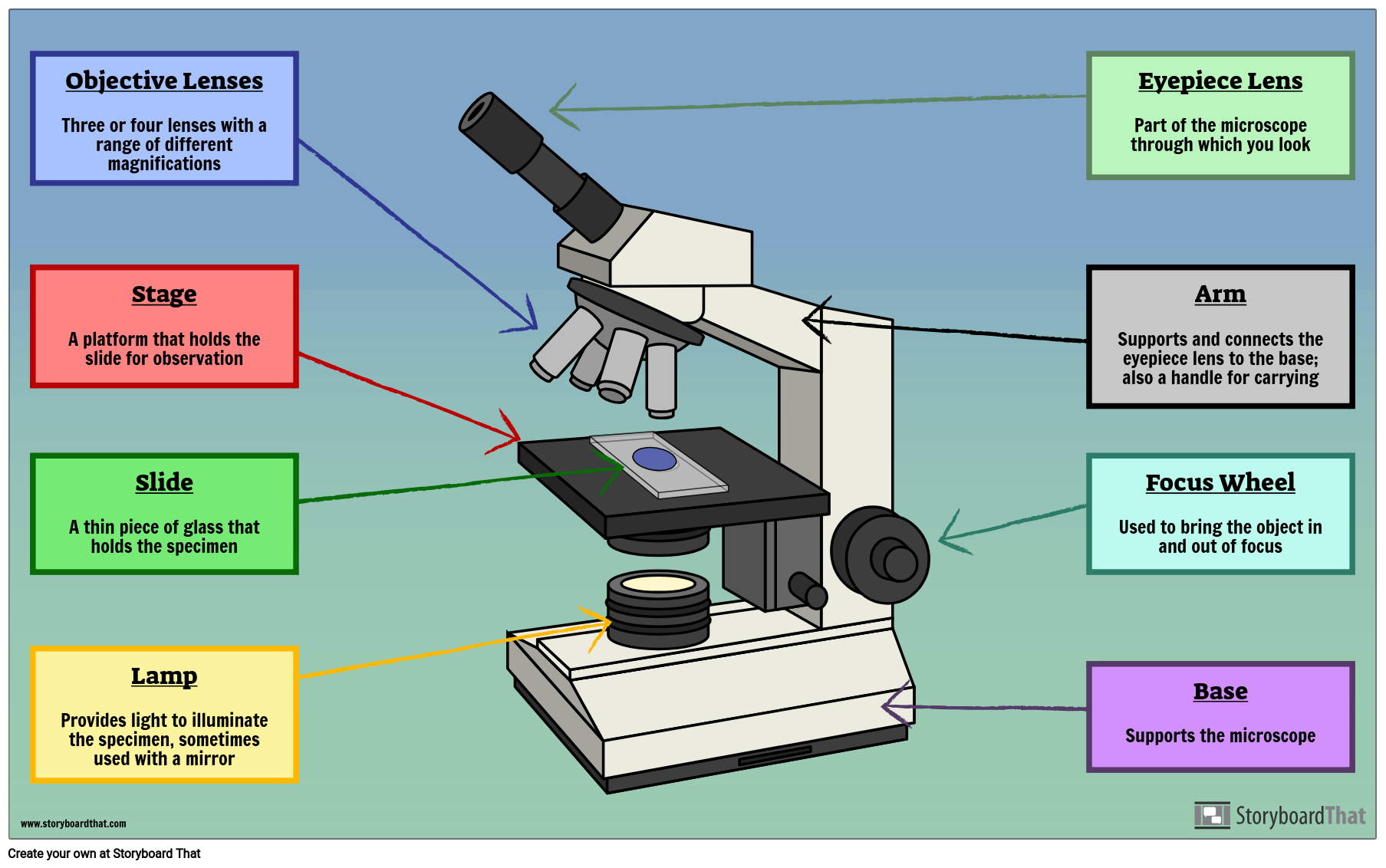
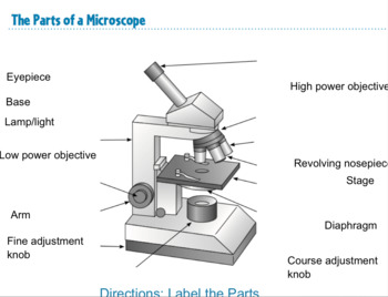
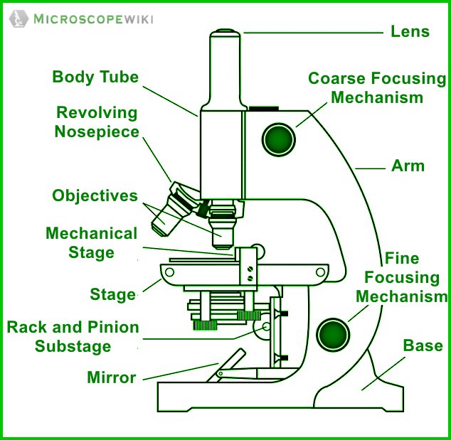
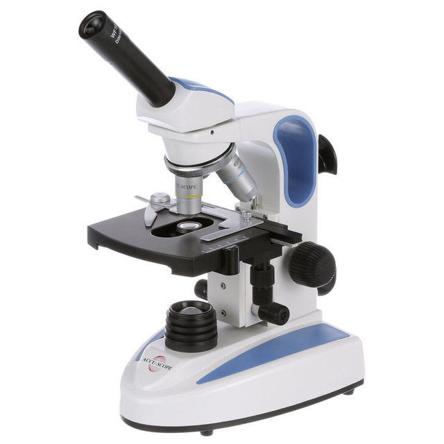
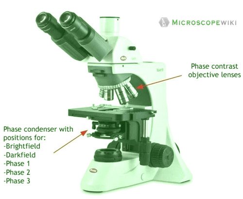
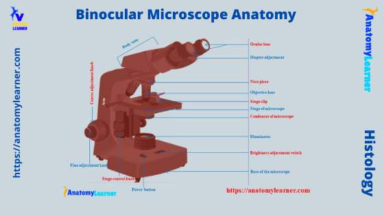
44 diagram of a microscope labeled
microbenotes.com › light-microscopeLight Microscope- Definition, Principle, Types, Parts ... Sep 7, 2022 · Parts of a microscope with functions and labeled diagram 22 Types of Spectroscopy with Definition, Principle, Steps, Uses History of Microbiology and Contributors in Microbiology rsscience.com › compound-microscope-parts-labeledCompound Microscope Parts – Labeled Diagram and their ... There are three major structural parts of a microscope: Head, Base, and Arm. Always lift a microscope by holding both the arm and base with two hands. There are two major optical lens parts of a microscope: Eyepiece (10x) and Objective lenses (4x, 10x, 40x, 100x).
microscopewiki.com › compound-microscopeCompound Microscope – Diagram (Parts labelled), Principle and ... Oct 10, 2022 · See: Labeled Diagram showing differences between compound and simple microscope parts. Structural Components. The three structural components include. 1. Head. This is the upper part of the microscope that houses the optical parts. 2. Arm . This part connects the head with the base and provides stability to the microscope.
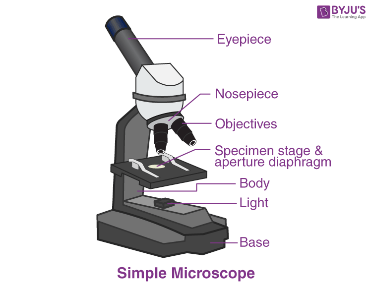
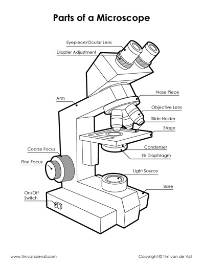
Diagram of a microscope labeled
anatomylearner.com › dog-skeleton-anatomyDog Skeleton Anatomy with Labeled Diagram - AnatomyLearner Dec 31, 2021 · Here, in the dog skeleton labeled diagram, I tried to show you the different segments of the forelimb, hindlimb with their bones. Again, I tried to show you all the bones from the vertebrae column of a dog skeleton. In addition, in the diagram, you will find a few identified skull bones. The sternum and the ribs are also identified in the dog ... anatomylearner.com › chicken-digestive-system-anatomyChicken Digestive System Anatomy with Labeled Diagram Dec 9, 2021 · The different parts of the small and large intestine of a chicken are showing below. I will discuss the anatomy of every single part of the small and large intestine with a labeled diagram. But, before going to intestine anatomy, I would like to introduce one of the exciting terms, gut-associated lymphatic tissue (GALT), with you. › createJoin LiveJournal Password requirements: 6 to 30 characters long; ASCII characters only (characters found on a standard US keyboard); must contain at least 4 different symbols;
Diagram of a microscope labeled. microbenotes.com › parts-of-a-microscopeParts of a microscope with functions and labeled diagram Sep 17, 2022 · Light Microscope- Definition, Principle, Types, Parts, Labeled Diagram, Magnification Amazing 27 Things Under The Microscope With Diagrams Plant Cell- Definition, Structure, Parts, Functions, Labeled Diagram › createJoin LiveJournal Password requirements: 6 to 30 characters long; ASCII characters only (characters found on a standard US keyboard); must contain at least 4 different symbols; anatomylearner.com › chicken-digestive-system-anatomyChicken Digestive System Anatomy with Labeled Diagram Dec 9, 2021 · The different parts of the small and large intestine of a chicken are showing below. I will discuss the anatomy of every single part of the small and large intestine with a labeled diagram. But, before going to intestine anatomy, I would like to introduce one of the exciting terms, gut-associated lymphatic tissue (GALT), with you. anatomylearner.com › dog-skeleton-anatomyDog Skeleton Anatomy with Labeled Diagram - AnatomyLearner Dec 31, 2021 · Here, in the dog skeleton labeled diagram, I tried to show you the different segments of the forelimb, hindlimb with their bones. Again, I tried to show you all the bones from the vertebrae column of a dog skeleton. In addition, in the diagram, you will find a few identified skull bones. The sternum and the ribs are also identified in the dog ...


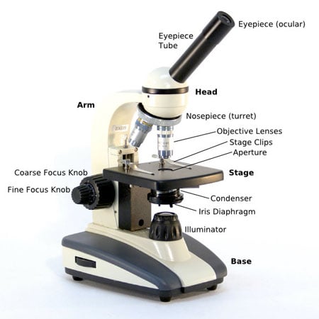
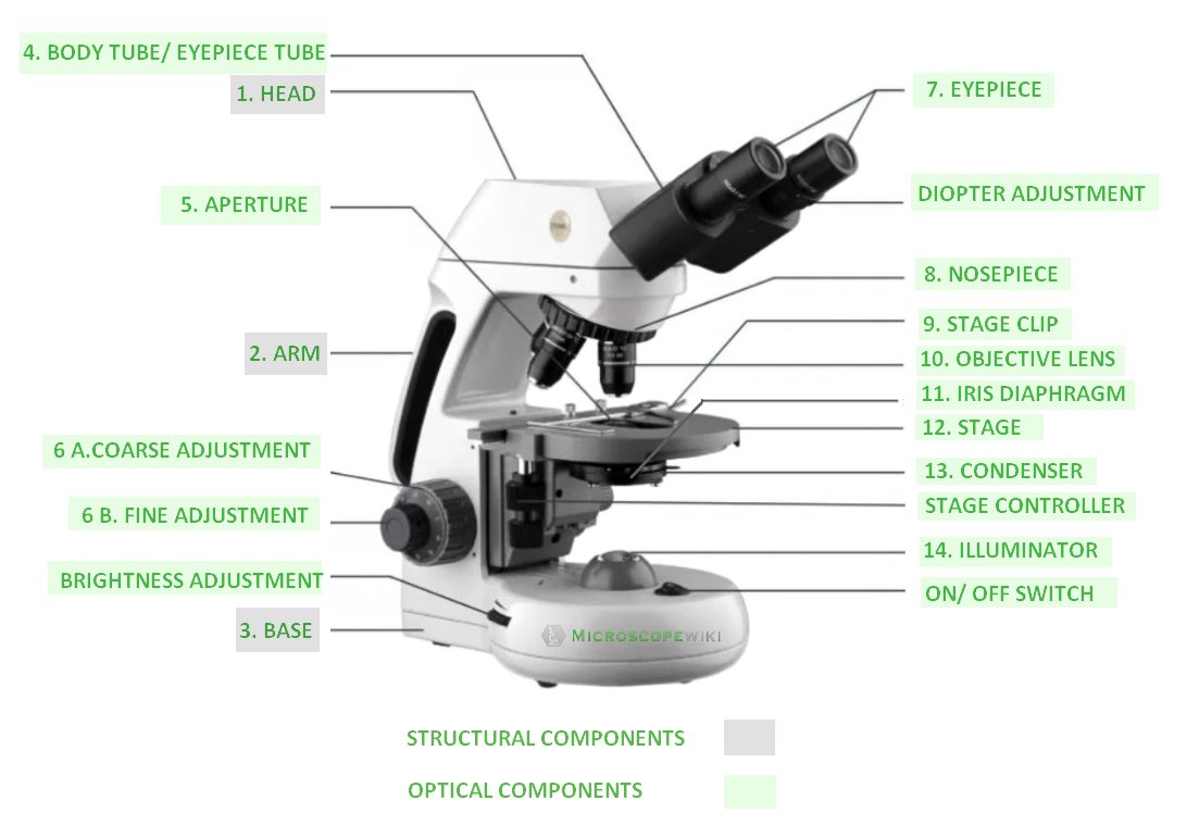













Post a Comment for "44 diagram of a microscope labeled"