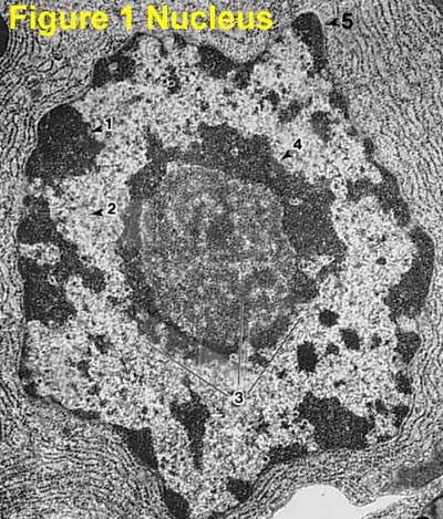
45 electron micrograph labeled
What Is the Adenovirus? - Healthline Apr 27, 2022 · The adenovirus is spread by close personal contact with others, like shaking hands or touching. Coughing, sneezing, or touching an object or surface that has the virus and then touching your mouth ... Join LiveJournal Password requirements: 6 to 30 characters long; ASCII characters only (characters found on a standard US keyboard); must contain at least 4 different symbols;
Scanning & Transmission Electron Microscopy Reveals Graphene ... Aug 20, 2021 · Figures 11 and 12 below shows a micrograph of different micro and nano particulates which have been identified in the Pfizer, Moderna, Astrazeneca and Janssen, so-called “vaccines” and analyzed under an Environmental Scanning Electron Microscope (SEM) coupled with an x-ray microprobe of an Energy Dispersive Spectroscopy (EDS) that reveals ...
Electron micrograph labeled
Electron microscope - Wikipedia An electron microscope is a microscope that uses a beam of accelerated electrons as a source of illumination. As the wavelength of an electron can be up to 100,000 times shorter than that of visible light photons, electron microscopes have a higher resolving power than light microscopes and can reveal the structure of smaller objects. A scanning transmission electron microscope … Bone and Bone Formation | histology - University of Michigan Slide 49 20x (humerus, H&E) View Virtual Slide Slide 49 40x (humerus, H&E) View Virtual Slide. There are two different magnifications (20X and 40X) of the epiphysis of a human long bone (those of you with even locker numbers may have a canine specimen on slide 49 that is much better). We require you to recognize 5 zones: 1) resting or reserve (R); 2) proliferative (P); 3) … Glossary - Molecular Biology of the Cell - NCBI Bookshelf Electron microscopy technique in which cellular structures or molecules of interest are labeled with antibodies tagged with electron-dense gold particles. These show up as black spots on the image. ... May be either a light micrograph or an electron micrograph depending on the type of microscope employed. microinjection. Injection of molecules ...
Electron micrograph labeled. Parvoviridae - Wikipedia Parvovirus virions are 23–28 nanometers (nm) in diameter and consist of the genome enclosed inside a capsid that is icosahedral in shape with a rugged surface. The capsid is composed of 60 structurally equivalent polypeptide chains derived from the C-terminal end of a VP protein's sequence, interlocking extensively to form an icosahedron with 60 asymmetric, superficial triangular units. Transmission Electron Microscopy - an overview | ScienceDirect … Hadis Rostamabadi, ... Seid Mahdi Jafari, in Characterization of Nanoencapsulated Food Ingredients, 2020. 2.5 Conclusion. Transmission electron microscopy (TEM) technique is a powerful tool for providing detailed information concerning the structural characteristics of nanoscale vehicles, e.g., nanoemulsions, nanoliposomal delivery systems, nanoparticles, nanofibers, etc. Transmission electron microscopy DNA sequencing - Wikipedia Transmission electron microscopy DNA sequencing is a single-molecule sequencing technology that uses transmission electron microscopy techniques. The method was conceived and developed in the 1960s and 70s, but lost favor when the extent of damage to the sample was recognized. In order for DNA to be clearly visualized under an electron microscope, it must be … Peripheral Nervous System | histology In this electron micrograph, note some of the features you saw in ventral horn motor neurons with the light microscope, such as the large, pale nucleus, prominent nucleolus, Nissl bodies, dendrites and axon. ... the discontinuous appearance of the axon labeled 5 in a Schwann cell is probably due to its curvature around the nucleus.
Glossary - Molecular Biology of the Cell - NCBI Bookshelf Electron microscopy technique in which cellular structures or molecules of interest are labeled with antibodies tagged with electron-dense gold particles. These show up as black spots on the image. ... May be either a light micrograph or an electron micrograph depending on the type of microscope employed. microinjection. Injection of molecules ... Bone and Bone Formation | histology - University of Michigan Slide 49 20x (humerus, H&E) View Virtual Slide Slide 49 40x (humerus, H&E) View Virtual Slide. There are two different magnifications (20X and 40X) of the epiphysis of a human long bone (those of you with even locker numbers may have a canine specimen on slide 49 that is much better). We require you to recognize 5 zones: 1) resting or reserve (R); 2) proliferative (P); 3) … Electron microscope - Wikipedia An electron microscope is a microscope that uses a beam of accelerated electrons as a source of illumination. As the wavelength of an electron can be up to 100,000 times shorter than that of visible light photons, electron microscopes have a higher resolving power than light microscopes and can reveal the structure of smaller objects. A scanning transmission electron microscope …
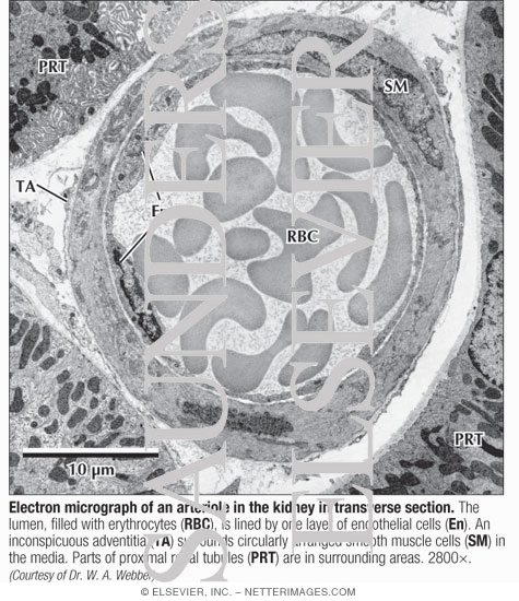
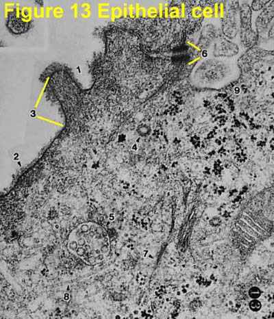
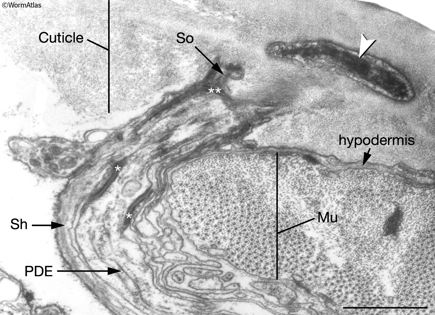
Electron Micrograph of an Arteriole In the Kidney In ...

Electron micrograph of section of a portion of rat glomerulus ...
cell and organelles Dr.Jastrow's electron microscopic atlas
Electron Micrographs

a Electron micrograph of rosette-forming lymphocyte, b ...
7,276 Electron Micrograph Stock Photos, Pictures & Royalty ...

Electron micrograph showing lack of mGluR1 labeling within a ...
Electron Micrographs
Transmission electron micrograph (TEM) identifying immunogold ...
Electron Micrographs

Electron micrograph of a labeled terminal with an 'apocrine ...
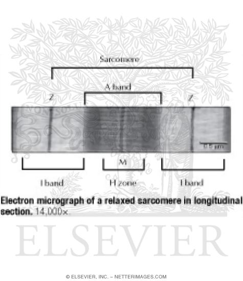
Electron Micrograph of a Relaxed Sarcomere In Longitudinal ...

Transmission electron micrograph of turkey spermatozoa ...
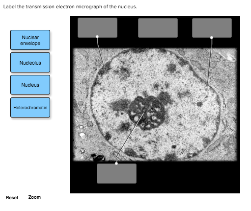
Solved Label the transmission electron micrograph of the ...

anatomy 10.png - Label the transmission electron micrograph ...
A tour of the cell: View as single page
animal cell electron micrograph labelling Diagram | Quizlet
2.3.3 Identify structures from electron micrographs of liver cells
Electron Micrographs
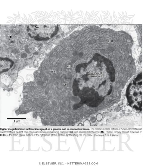
Higher Magnification Electron Micrograph of a Plasma Cell In ...

Freeze-fracture electron micrograph of spinach thylakoids. a ...
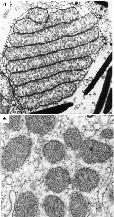
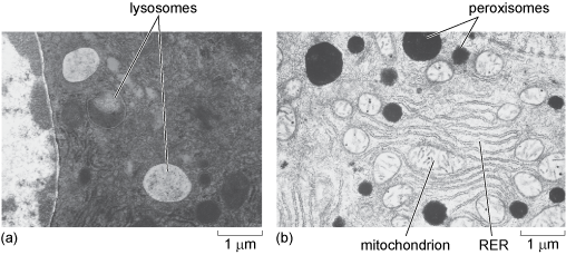
Transmission electron micrographs of mitochondria, site of ...
Electron Micrographs
Electron Micrograph of Actin and Intermediate Filaments In ...
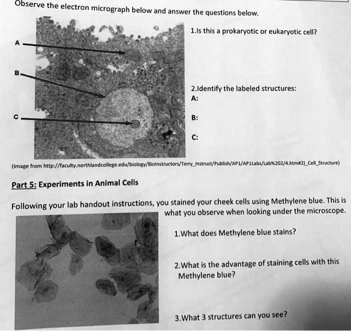
SOLVED: Observe the electron micrograph below and answer the ...
A tour of the cell: View as single page
ExcFIG 4 Legend
Electron Micrographs
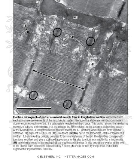
Electron Micrograph of Part of A Skeletal Muscle Fiber In ...
CcFIG 5 Legend
Electron Micrographs

2.2.3 Identify structures in electron micrographs of Ecoli ...
A and B) Electron micrograph of a cell labeled for/5-tubulin ...
cell and organelles Dr.Jastrow's electron microscopic atlas
IntroFIG 4B Legend
Electron micrographs from a nonimmunogold-labeled section (A ...
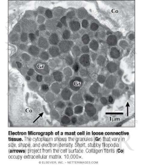
Electron Micrograph of a Mast Cell In Loose Connective Tissue

Transmission electron micrograph of a gold-labelled Lowicryl ...
Solved label the ectron micrograph of an animal cell. | Chegg.com

Transmission electron micrograph of sodium phosphotungstate ...

Transmission electron micrograph of a thin section through ...

Label electron micrograph of B lymphocyte. - Brainly.com
Electron Micrographs

Electron micrographs of SPIO-labeled MSCs. A, Cell nucleus (N ...
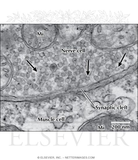
Electron Micrograph of Synaptic Vesicles at a Neuromuscular ...
Post a Comment for "45 electron micrograph labeled"