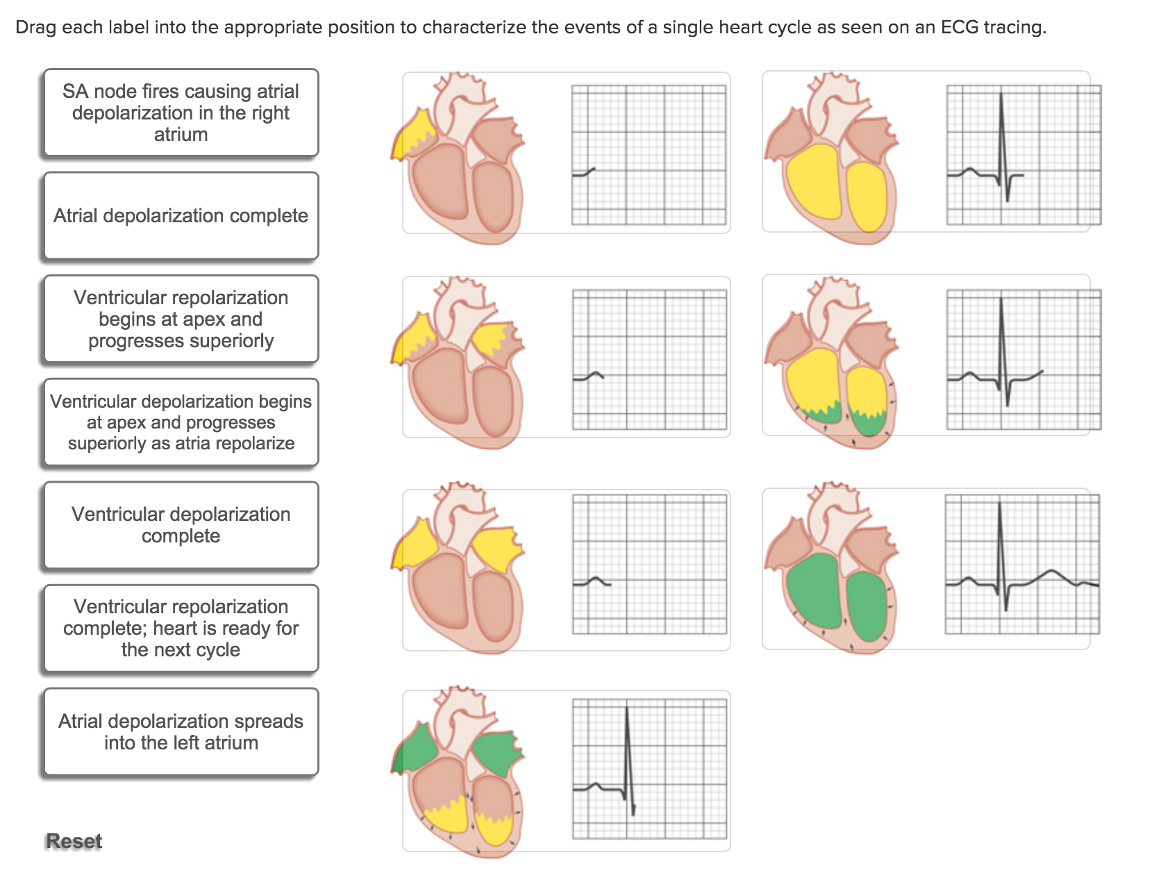
42 drag each label into the appropriate position to characterize the events of a single heart cycle
Join LiveJournal Password requirements: 6 to 30 characters long; ASCII characters only (characters found on a standard US keyboard); must contain at least 4 different symbols; [Solved] Drag each label into the appropriate position to characterize ... Drag each label into the appropriate position to characterize the events of a single heart cycle as seen on an ECG tracing. SA node fires causing atrial depolarization in the right atrium Atrial depolarization complete Ventricular repolarization ... Show more ... Show more Biology Science Anatomy BIO 211 Answer & Explanation
(PDF) B Research Methods ForBus A Skill Building Approach ... B Research Methods ForBus A Skill Building Approach 7e2016UmaSekaran, RogerBougie Wiley

Drag each label into the appropriate position to characterize the events of a single heart cycle
An Introduction to Mechanics Kleppner Kolenkow 2e Enter the email address you signed up with and we'll email you a reset link. Cell Cycle - Definition And Phases of Cell Cycle - BYJUS Phases of Cell Cycle Cell cycle or cell division refers to the series of events that take place in a cell leading to its maturity and subsequent division. These events include duplication of its genome and synthesis of the cell organelles followed by division of the cytoplasm. Solved Drag each label into the appropriate position to - Chegg Drag each label into the appropriate position to characterize the events of a single heart cycle as seen on an ECG tracing. Show transcribed image text Expert Answer 100% (23 ratings) Figure 1 SA node fires causing atrial depolarization in right atrium Figure 5 ventricular de … View the full answer
Drag each label into the appropriate position to characterize the events of a single heart cycle. Chapter 15 Cardiovascular Practice Flashcards | Quizlet Complete each sentence, and then place them in the correct order to describe blood flow through the heart, beginning with blood entering the right side of the heart. Beginning with the return from the systemic circulation, blood enters the right atrium. Blood then travels through the tricuspid valve and into the right ventricle. Solved Drag each label into the appropriate position to - Chegg transcribed image text: chapter 19 worksheet g seved help save & exit submit chapter 19 worksheet drag each label into the appropriate position to characterize the events of a single heart cycle as seen on an ekg tracing 2 ventricular begins at the atrial apex and 0.27 points superiorly ass completed repolarize ventricular repolarization and the … Chpt. 1, 3, 24, and 9 HW - Weebly a voltage or electrical charge across the plasma membrane. 9. During interphase of the cell life cycle, the cell divides into two cells. False. 10. In their resting state, all body cells exhibit a resting membrane potential; therefore, all cells are polarized. ALEX | Alabama Learning Exchange Students will change a variable such as: number of students riding the snow sled, size of the child (children) riding the snow sled, direction, position on the hill the snow sled is released, position of children on the sled (sitting, standing, laying), friction caused by materials that makes up the sled, and air resistance caused by an object such as a parachute. Students will collect and ...
CV Physiology | Cardiac Cycle Diastole represents the period of time when the ventricles are relaxed (not contracting).Throughout most of this period, blood is passively flowing from the left atrium (LA) and right atrium (RA) into the left ventricle (LV) and right ventricle (RV), respectively (see figure at right). Heart Diagram with Labels and Detailed Explanation - BYJUS Diagram of Heart. The human heart is the most crucial organ of the human body. It pumps blood from the heart to different parts of the body and back to the heart. The most common heart attack symptoms or warning signs are chest pain, breathlessness, nausea, sweating etc. The diagram of heart is beneficial for Class 10 and 12 and is frequently ... Solved Drag each label into the appropriate position to - Chegg question: drag each label into the appropriate position to characterize the events of a single heart cycle as seen on an ekg tracing ventricular ventricular depolarization repolarization begins at the is complete and the heart apex and progresses is ready for the superiorly as next cycle. atria repolanze atrial ventricular depolarization … Solved Drag each label into the appropriate position to - Chegg Transcribed image text: Drag each label into the appropriate position to characterize the events of a single heart cycle as seen on an EKG tracing.
Circulatory System: Blood Flow Pathway Through the Heart Left Ventricle & Right Ventricle. The ventricles are the two lower chambers of the heart.The right ventricle receives oxygen-poor blood from the right atrium and pumps it through the pulmonic semilunar valve to the pulmonary artery and into the lungs to be filled with oxygen. On the other hand, the left ventricle receives oxygen-rich blood from the left atrium and pumps it through the aortic ... Drag each tile to the correct box. Not all tiles will be used. The ... Drag each tile to the correct box. Not all tiles will be used. The basic components of an ex nihilo creation story are below. The first sentence of the story is provided. Complete the ex nihilo story by putting the correct sentences in the proper order. A long time ago, there was nothing but darkness. The animal dived into the water and brought ... Heart Lecture Flashcards | Quizlet Correctly label the following external anatomy of the posterior heart. Drag each label into the appropriate position to characterize the events of a single heart cycle as seen on an EKG tracing. Correctly label the pathway of blood flow through the heart, beginning with the right atrium. Recommended textbook solutions 38 Drag each label into the appropriate position to characterize the ... 38.Drag each label into the appropriate position to characterize the events of a single heart cycle as seen on an ECG tracing. Note: focus on the black area of each part of the ECG.
PDF In this chapter, you will learn that - Pearson The endomysium(en″do-mis′e-um; "within the muscle") is a wispy sheath of connective tissue that sur- rounds each individual muscle fiber. It consists of fine areo- lar connective tissue. As shown in Figure 9.1, all of these connective tissue sheaths are continuous with one another as well as with the tendons that join muscles to bones.
PDF The Cardiovascular System - Pearson tomical areas of the heart are described in the next section, keep referring to Figure 11.3 to locate each of the heart structures or regions.) Chambers and Associated Great Vessels Learning Objectives Trace the pathway of blood through the heart. Compare the pulmonary and systemic circuits. The heart has four hollow cavities, or chambers—
Labeling, ranking, sorting, or sentence completion questions All of these question types require you to position items into an area of the answer box. Answer these kinds of questions on a computer, not on a smartphone. Basic keyboard instructions (applies to most keyboards) Labeling questions. Ranking questions. Sorting questions. Sentence completion (vocabulary) questions.
Chapter 19: The Heart Flashcards | Quizlet Size, Shape and Position of the Heart •Located in thoracic cavity -specifically in the mediastinum •area between lungs -superior to diaphragm -posterior to sternum -2/3 of heart to the left of midsagittal plane due to the liver taking space on the right •Base - broad superior portion •Apex - inferior end, tilts to the left, tapers to point
Answered: Muscle cells, neurons, and red blood… | bartleby Q: Drag each label into the appropriate position to identify whether the statement or image depicts… A: There are four types of connective tissues in the human body that is bone, blood, connective tissue…
[Expert Answer] Drag the tiles to the boxes to form correct pairs ... Drag the tiles to the boxes to form correct pairs. Match each state of matter with the statement that best describes it. plasma Particles move past each other freely but do not go far apart. gas Particles are so hot that electrons are stripped from atoms. liquid It retains its shape regardless of the shape of the container. solid It expands to fill the volume of the container.
Cardiac Cycle- Physiology, Diagram, Phases of the Cardiac Cycle - BYJUS In a normal person, a heartbeat is 72 beats/minute. So, the duration of one cardiac cycle can be calculated as: 1/72 beats/minute=.0139 minutes/beat. At a heartbeat 72 beats/minute, duration of each cardiac cycle will be 0.8 seconds. Duration of different stages of the cardiac cycle is given below: Atrial systole: continues for about 0.1 seconds
Ch. 19 Circulatory System- heart Flashcards | Quizlet Correctly label the external anatomy of the anterior heart. Place the labels in order denoting the flow of blood through the pulmonary circuit beginning with the right atrium and ending in the left atrioventricular valve. The first and last structures are given. Right atrium 1. tricuspid valve 2. right ventricle 3. pulmonary valve
Chapter 19,20,21 Flashcards | Quizlet Drag each label into the appropriate position to characterize the events of a single heart cycle as seen on an ECG tracing. Drag each statement to the appropriate position to identify the valve being described. The __________ valve is between the right atrium and right ventricle. Tricuspid Select all that are true regarding ventricular balance. 2,4
Solved Drag each label into the appropriate position to - Chegg Expert Answer. Parts of Normal ECG: 1. P wave: It denotes atrial depolarization. 2. QRS Complex: This is caused by ventricular depolarization. 3. Q wave: If the first wave of …. View the full answer. Transcribed image text: Drag each label into the appropriate position to identify the waves of a normal ECG Alpha wave 0 QRS complex ST segment ...
Phases of the Cardiac Cycle When the Heart Beats - ThoughtCo The cardiac cycle is the sequence of events that occurs when the heart beats. As the heart beats, it circulates blood through pulmonary and systemic circuits of the body. There are two phases of the cardiac cycle: The diastole phase and the systole phase. In the diastole phase, heart ventricles relax and the heart fills with blood.
Question 5 1 1 point which of the following - Course Hero Pages 7 ; Ratings 100% (12) 12 out of 12 people found this document helpful; This preview shows page 2 - 5 out of 7 pages.preview shows page 2 - 5 out of 7 pages.
Cardiac cycle phases: Definition, systole and diastole | Kenhub The cardiac cycle is defined as a sequence of alternating contraction and relaxation of the atria and ventricles in order to pump blood throughout the body. It starts at the beginning of one heartbeat and ends at the beginning of another. The process begins as early as the 4th gestational week when the heart first begins contracting.
Select all that are true regarding potassium balance. A-The... Q: Drag each label into the appropriate position to characterize the events of a single heart cycle as seen on an ECG traci Q: The nurse practitioner is performing a thoracentesis on a patient with a pleural effusion.
Cardiac Cycle Phases and Blood Flow: Step-By-Step Heart Diagram - EZmed There are 2 main phases to the heart cycle - diastole and systole. Diastole is defined as the phase in which the heart, especially the ventricles, is at rest. The relaxed heart allows for blood to fill the cardiac chambers. Systole is defined as the phase in which the heart, especially the ventricles, is contracting.
Solved Drag each label into the appropriate position to - Chegg Drag each label into the appropriate position to characterize the events of a single heart cycle as seen on an ECG tracing. Show transcribed image text Expert Answer 100% (23 ratings) Figure 1 SA node fires causing atrial depolarization in right atrium Figure 5 ventricular de … View the full answer
Cell Cycle - Definition And Phases of Cell Cycle - BYJUS Phases of Cell Cycle Cell cycle or cell division refers to the series of events that take place in a cell leading to its maturity and subsequent division. These events include duplication of its genome and synthesis of the cell organelles followed by division of the cytoplasm.
An Introduction to Mechanics Kleppner Kolenkow 2e Enter the email address you signed up with and we'll email you a reset link.










Post a Comment for "42 drag each label into the appropriate position to characterize the events of a single heart cycle"